Home
/ Animal Cell Diagram With Labels And Functions : A Labeled Diagram Of The Animal Cell And Its Organelles Biology Wise / It also organizes some of the cell components maintaining the cell shape 4.
Animal Cell Diagram With Labels And Functions : A Labeled Diagram Of The Animal Cell And Its Organelles Biology Wise / It also organizes some of the cell components maintaining the cell shape 4.
Animal Cell Diagram With Labels And Functions : A Labeled Diagram Of The Animal Cell And Its Organelles Biology Wise / It also organizes some of the cell components maintaining the cell shape 4.. Within its membranes, there are membranous spaces called the cristae spaces and the membrane folding are called cristae. The outer membrane is permeable, allowing t. On the ribosomes, the mrna helps determine the coding for transfer rna (trna) which also determines the protein amino acid sequences. It also organizes some of the cell components maintaining the cell shape 4. It is the site for protein synthesis.
A single replicated cell has about 10 million ribosomes. The number of mitochondria found in each cell varies widely depending on the function of the cell it performs. Cell membrane or plasma membrane is a membrane common to both plant and animal cells. The animal cell diagram is widely asked in class 10 and 12 examinations and is beneficial to understand the structure and functions of an animal. The working together of all cells gives an animal its ability to move, to reproduce, to respond to stimuli, to digest and absorb food, etc.
What Are The Parts Of An Animal Cell And Its Functions Quora from qph.fs.quoracdn.net This is a continuous folded membranous organelle found in the cytoplasm made up of a thin network of flattened interconnected compartments (sacs) that connects from the cytoplasm to the cell nucleus. Manufacturing, processing and transporting proteins for cell utilization both in and out of the cell. Rough er transports the proteins and lipids through the cell into the cristae. The function of the ribosomes on rough er is to synthesis proteins and they have a signaling sequence, directing them to the endoplasmic reticulum for processing. These subunits are designated as the 40s and 60s in the animal cell. Generally, the combined effort by all animal cells is what enables the normal functioning of the body. Feb 27, 2018 · an animal cell diagram is a great way to learn and understand the many functions of an animal cell. It also organizes some of the cell components maintaining the cell shape 4.
Within its membranes, there are membranous spaces called the cristae spaces and the membrane folding are called cristae.
Generally, the combined effort by all animal cells is what enables the normal functioning of the body. The cytoskeleton functions to create a network organizing the cell components and to also maintain the cell shape. A single replicated cell has about 10 million ribosomes. More images for animal cell diagram with labels and functions » See full list on microbenotes.com See full list on microbenotes.com Each ribosome is made up of two subunits i. The nerves and muscles are made up of specialized cells that plant cells. The cell is the structural and functional unit of life. See full list on microbenotes.com The function of the ribosomes on rough er is to synthesis proteins and they have a signaling sequence, directing them to the endoplasmic reticulum for processing. In a eukaryotic cell, ribosomes constitute half ribosomal rna and half ribosomal proteins. The membrane is selectively permeable and allows only certain molecules to pass through.
See full list on microbenotes.com These subunits are designated as the 40s and 60s in the animal cell. As observed in the labeled animal cell diagram, the cell membrane forms the confining factor of the cell, that is it envelopes the cell constituents together and gives the cell its shape, form, and existence. Within its membranes, there are membranous spaces called the cristae spaces and the membrane folding are called cristae. Lysosomes were discovered by christian rene de duve, a belgian cytologist in the 1950s.
Draw A Well Labeled Diagram Of Animal Cell Home Work Help Learn Cbse Forum from ask.learncbse.in Helping in cell division by allowing separation of chromosomes Manufacturing, processing and transporting proteins for cell utilization both in and out of the cell. The cytoskeleton functions to create a network organizing the cell components and to also maintain the cell shape. See full list on microbenotes.com Listed below are the cell organelles of an animal cell along with their functions. On the ribosomes, the mrna helps determine the coding for transfer rna (trna) which also determines the protein amino acid sequences. The nucleus and its component organelles are suspended in the nucleoplasm (house of the chromosomal dna and genetic materials) Rer rough endoplasmic reticulum synthesizes proteins for secretion membrane proteins and organelle proteins.
Listed below are the cell organelles of an animal cell along with their functions.
See full list on microbenotes.com A cell has one nucleus which divides producing multinucleated cells e.g. The membranes bend into folds known as cristae. Cytosol is the fluid present within a cell that is made up of water and ions such as potassium, proteins and small molecules. These proteins are found in the cell cytoplasm of the eukaryotic cells. The cell wall is present: The ribosomal subunits are the site for genetic coding into proteins. The er has more than half the membranous cell content, hence it has a large surface area where chemical reactions take place. It holds other cells organelles including the nucleolus, nucleosomes, and chromatins. It is held together to the cytoplasm with the help of the filaments and microtubules. They are also found in cilia and flagella. Therefore, the nucleus is the information center. It also organizes some of the cell components maintaining the cell shape 4.
Apr 26, 2021 · plant cell: These proteins are found in the cell cytoplasm of the eukaryotic cells. See full list on microbenotes.com On the ribosomes, the mrna helps determine the coding for transfer rna (trna) which also determines the protein amino acid sequences. The cell wall is absent:
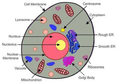
What Is Animal Cell Its Structure And Function With Diagram Micro B Life from i1.wp.com As observed in the labeled animal cell diagram, the cell membrane forms the confining factor of the cell, that is it envelopes the cell constituents together and gives the cell its shape, form, and existence. Diagram of animal cell, created with biorender.com It also organizes some of the cell components maintaining the cell shape 4. Manufacturing, processing and transporting proteins for cell utilization both in and out of the cell. The primary role of the nucleus is to control and regulate cell activities of growth and maintain cell metabolisms. It is the site for protein synthesis. It also carries the genes that have hereditary information of the cell. Ribosomes that occur as free particles are attached to the endoplasmic reticulum membrane occurring in large numbers accounting for about a quarter of the cell organelles.
The cell is the structural and functional unit of life.
They are then sent into the golgi bodies or inserted into the cell membrane. This is a fibrous network that's formed from and by different proteins of long chains of amino acids. More images for animal cell diagram with labels and functions » Centrioles are about 500nm long and 200nm in width that are found close to the nucleus and helps in cell division. The outer membrane is permeable, allowing t. All living cells contain ribosomes, which may be freely circulating in the cytoplasm and some are bound to the endoplasmic reticulum. Some cells lose their nuclei after maturati. Ribosomes that occur as free particles are attached to the endoplasmic reticulum membrane occurring in large numbers accounting for about a quarter of the cell organelles. Feb 27, 2018 · an animal cell diagram is a great way to learn and understand the many functions of an animal cell. It also provided a uniform movement of the cell and its organelles, by the filament system network found in the cell's cytoplasm. See full list on microbenotes.com The chromosomal dna and genetic materials, which are made up of genetic coded ultimately make up their proteins' amino acid sequences for use by the cell. Each ribosome is made up of two subunits i.
Share :
Post a Comment
for "Animal Cell Diagram With Labels And Functions : A Labeled Diagram Of The Animal Cell And Its Organelles Biology Wise / It also organizes some of the cell components maintaining the cell shape 4."


Post a Comment for "Animal Cell Diagram With Labels And Functions : A Labeled Diagram Of The Animal Cell And Its Organelles Biology Wise / It also organizes some of the cell components maintaining the cell shape 4."